COMP-Northwest (OR): Medical Anatomy Center
Medical Anatomy Center Vision
The vision of the Medical Anatomy Center (MAC) is to advance the teaching, learning and research of medical knowledge, patient & community care by integrating dynamic anatomy, innovation, imaging, and emerging technologies. To further develop physical examination/palpation/manual treatments, ultrasound, clinical skills, and surgery while developing quality simulation producing today’s safe efficient physician, who will address prevention, early diagnosis, rehabilitation, and maintenance, enabling a platform for future improvements in medicine producing healthier communities for global sharing. The MAC’s vision is implemented through serving learners, institutions, and healthcare.
- First and foremost, the Medical Anatomy Center serves to improve and integrate healthcare student skills and experiences, advancement of medicine through education and research and contribute to safe, quality care to individuals, local communities, and global health.
- Promote healthcare fundamentals with dynamic anatomy, integrating clinical acumen, patient experiences and innovation between academia, private industry, and communities.
- Demonstrate excellence in teaching and learning dynamic, medically relevant anatomy through best practices including ratio anatomy values, variation, and diversity.
- Integrate medical dynamic anatomy and physical examination with ultrasound imaging as a basis for assessing health, clinical diagnosis, osteopathic treatments while promoting the safe delivery of health & care, disease prevention and/or maintenance to empower an individual to define their own quality of life as a member of a community.
- Teach medical students/trainees the biomedical research process, teamwork, and professionalism to advance evidence-based medicine.
- Be leaders in medical education through observation, disruptive innovation, emerging technologies, passion, and humanism, enhancing learning and teaching philosophies, methods, and techniques to improve the health and knowledge of individuals and communities.
- Lead and support the medical profession in advancing the understanding and value of ever evolving dynamic anatomy in medical education, research, technology, patient care and global health.
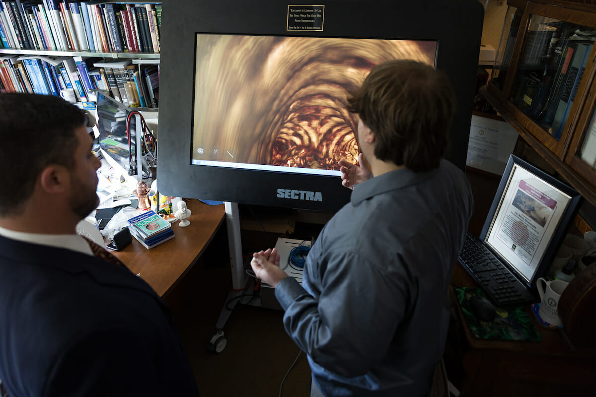
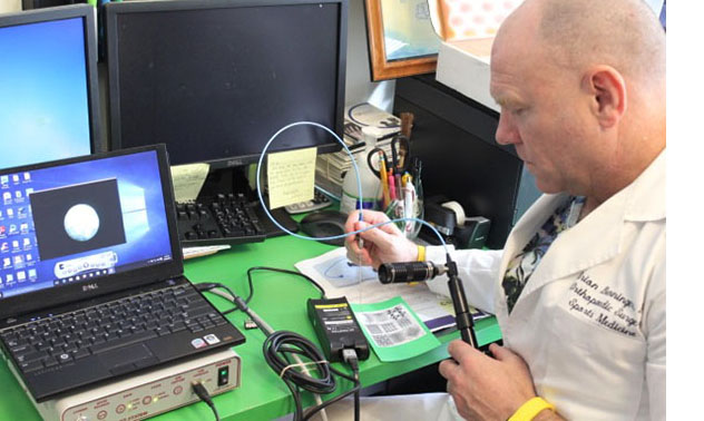
 Dr. Brion Benninger (Sports Medicine, Clinical Anatomist, Reverse Translational Researcher and Innovative Medical Educator) is the Executive Director of the Medical Anatomy Center, Western University of Health Sciences, Lebanon, Oregon and faculty to the General Surgery, Orthopedic and Sports Medicine residency programs at Samaritan Health Services, Corvallis Oregon. Dr. Benninger is known internationally as one of today’s most progressive medical educators and his work with Google Glass has lead him to be on the Google Glass Northwest Development Team. He has taught and worked in the medical field in several countries, which has provided him invaluable insight and experience. The Lebanon community engagement and support is such an important part of the culture our medical students experience during their time at this unique osteopathic medical school (COMP-Northwest site). Dr Benninger continues to thank the Lebanon community for so fully embracing our students and supporting them as they strive to become the doctors of the future. He has mentored over 100 students on research projects that have been presented at national and international meetings and his students have received several research awards from conferences, but he is most proud of the overwhelming comments he receives regarding the students’ professionalism. “I am pleased that our students represent the medical school from WesternU and the community of Lebanon, Oregon with such pride. I would also like to thank Dr. Paula Crone, Medical Dean of WesternU, for supporting these innovative teaching methods to further enrich the experience of our students at COMP-Northwest and WesternU.” Dr. Benninger believes recognizing and teaching people with different strengths to become effective, caring, safe, and efficient physicians must be viewed as a moving target requiring different weapons for success. This starts with recognizing the different learning strengths people have and providing them the opportunity to flourish. It needs an infrastructure which encourages looking outside contemporary medical education and being willing to develop innovative ideas. To many of today’s established institutions, teaching tried and tested pearls and theories regardless of the topic or technique is considered wise. To take a risk and challenge tried and tested methods using technology, however, can be viewed as cutting corners. Dr. Benninger’s goal, at the risk of being labeled “unconventional”, is to constantly challenge how current education systems teach healthcare students to become caring, safe, and efficient providers and to push the utilization of new technologies at the educational level. Integrating technology, human traits and behavior creates and delivers disruptive innovative education and research. I have witnessed and analyzed the positives and what needs improved from both allopathic and osteopathic medical schools of training. One of the issues in healthcare education at Academic institutions is that the curriculum is based on pedagogical (children) learning theories which have been successful in educating children but are not based on andragogical (adult) theories for today’s adult learner. The average adult can focus for 12-18 minutes, then needs different stimulation or a break before returning to the topic at hand. It has been shown that playing music while attempting a fine detailed task (whether it’s surgery or fine sculpting) reveals better results. These facts are why Dr. Benninger promotes a technique teaching in multiples of 20-minute intervals and encourages listening to music while dissecting or conducting invasive skills in the anatomy lab. Some students learn more effectively with movement and integration, performing physical exams on each other while simultaneously conducting ultrasound addressing surface and deep anatomy. When performing several exams on each other and on patients, they add palpation skills including skin surface tension, temperature, moisture, muscle tone, proprioception and physiological end points into muscle and other memory banks. Simultaneously using a novel ultrasound finger probe by Sonivate (which he helped with research and development) during the examination increases their ultrasound probe time and stereostructural anatomy identification skills. The combination of these activities causes signals to enter the students’ nervous system, which are packaged as memory and can be retrieved efficiently similar to a fine tuned athlete performing a task. These are direct benefits of using “unconventional” ways, supported by research, to teach people with different strengths to become effective, caring, safe, and efficient physicians. A successful physician or healthcare provider applies the art and science of medicine to maladies affecting the social, psychological and physical aspects of humans, and that is exactly what we are striving to teach our students. Unconventional tactics also lead us to new discoveries and new techniques. During the closing events of the International Association of Medical Science Educators in St Andrews Scotland, Dr. Benninger was referred to as “a genius” for his multiple innovative education approaches using emerging technologies to integrate basic science and clinical skills simultaneously. He simply wants to address as many strengths and styles as possible. He successfully integrated Google Glass and other Optical Head Display units into the medical anatomy lab curriculum while conducting several pilot studies, which were firsts with Google Glass. These pilots resulted in his development of the Triple Feedback Examination technique by integrating Google Glass, ultrasound and a linear finger probe (Fukuda-Denshi & SonicEye respectively) creating triple feedback during physical examinations. The physician’s hand is a palpation stethoscope, the ultrasound finger probe is a visual stethoscope, and integrating them with Glass provides a physician with 3 forms of feedback during physical examinations (palpation, surface, and internal visualization). The Triple Feedback Examination technique incorporates human touch, physical movement with muscle memory, surface and deep anatomy while making eye contact with the patient using Google Glass. Dr. Benninger has delivered several presentations worldwide regarding his signature Triple Feedback Examination Technique and how reinventing old techniques with innovative technology has large implications for both civilian and military medicine. He was the first to use the Triple Feedback Examination technique to successfully identify shrapnel that can be lodged in upper and lower limbs during military combat and civilian firearm violence, which will lead to quicker & safer emergency surgeries of soldiers and police officers. He was also the first to use the Triple Feedback Examination technique to prove that images can be successfully obtained from donor cadavers and live subjects, which will improve our training methods across many medical fields. Was the first to use the Triple Feedback Examination technique to identify fractures of the mandible on donor cadaver patients. First to combine Google Glass with the DirectVision urinary cathether by PercuVision, which allowed viewing the urethral contents through the Glass while placing a urinary catheter, which will ultimately diminish the trauma inflicted from blind catheter placement and improve the quality of care to patients. Not only is his blending of emerging technologies with new techniques improving medicine, but his advances are changing the way we teach as well. In July of 2014, he was the first to successfully assess medical students anatomy knowledge using Google Glass during anatomy lab examination, identifying cadaver structures without the traditional use of pins, flags and strings, removing structures biases and representing anatomy as it would be experienced during physical examinations and invasive techniques. In August of 2014, he was the first to deliver a formal radiology lecture to first year medical students using Google Glass. In September of 2014, he was the first to teach medical students how to place urinary catheters using DirectVision on an advanced urinary simulator and then on donor cadavers using Google Glass. In October of 2014, he was the first to combine Google Glass with ultrasound linear finger probe during a Procedures Lab for Emergency Medicine, Military, Surgical, Ultrasound, Othopaedic and Sports Medicine Clubs attended by first and second year medical students at COMP-Northwest in Lebanon Oregon and conducted by Dr. Eschelbach, Medical Director at St Charles Health System, Redmond Oregon. Students were able to conduct and practice the skills of the Seldinger technique for central lines by combining Google Glass and SonicEye finger ultrasound linear transducer with a Fukuda-Denshi ultrasound system. Dr. Benninger believes it can be considered reckless or lazy to break from the tried & tested traditions of our old medical techniques, but unconventional methods provide the new discoveries that drive our innovation. Our successes with our multiple education approaches and integrating emerging technologies such as Google Glass and the SonicEye finger ultrasound linear & biplanar transducers speak for themselves. Dr. Benninger is proud to be working with medical students to test & develop new techniques taking advantage of emerging technologies, and proud of how our successes are representing COMP-Northwest & COMP across the world. Lebanon’s connection to COMP-Northwest has cultivated a support system and a culture, which has fostered a willingness to challenge old ways that has been embraced by Dean Crone and staff. This willingness to break away from the status quo is a large part of what makes COMP-Northwest & COMP one of the most unique and leading-edge medical colleges. With the unique blend between our medical school and caring community and teaching students with disruptive innovation, it will assure effective, caring, safe, and efficient physicians of the future.
Student Research Program
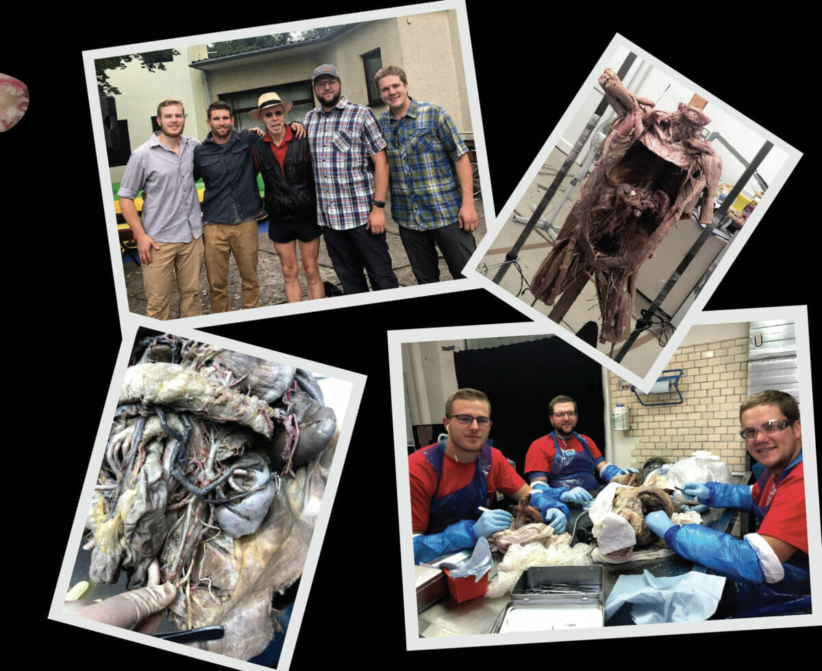
- Integrating innovation and technology, developing student research projects which teach the research process leading to oral/poster presentation skills for National/International conferences and publications.
- Research areas include: medical education, reverse translational research, clinical science, emerging technologies, disruptive innovation, ultrasound education including anatomy, invasive procedures, maintenance of chronic injuries and physical examination, imaging and stereostructural anatomy, military medicine, concussion/sports medicine, hyperbaric medicine, surgical radiological and anatomical terminology and history of medicine.
Continuing Medical Education Program
Conduct CME courses related to radiology, invasive procedures and clinical anatomy
Clinical Integration Program
Teach the utility of anatomical knowledge as a platform for diagnosis at all levels of medical experience.
Visiting Professor Program
Collaborate with world expert transitional researchers, physicians and educators.


